
Retroperitoneal Sarcoma treatment in Bangalore requires specialist evaluation because these rare tumors often lie close to the kidneys, bowel, pancreas, nerves, and major blood vessels. Dr Suraj Manjunath, a senior Surgical Oncologist at Apollo Hospitals, evaluates the tumor’s type, extent, and relationship with nearby organs to plan safe, complete removal and coordinate additional treatment when required.


The retroperitoneum is an area at the back of the abdomen. It contains or surrounds structures such as the kidneys, adrenal glands, ureters, pancreas, portions of the bowel, muscles, nerves, and major blood vessels.
A retroperitoneal sarcoma begins in the soft tissues within this space. Sarcomas are different from the more common cancers that begin inside organs. They arise from tissues such as fat, muscle, fibrous tissue, blood vessels, or nerves.
The common types include:
The subtype matters because different sarcomas behave differently. Some are more likely to return in the abdomen, while others have a greater tendency to spread to distant organs. The surgical strategy may also differ between a liposarcoma and a leiomyosarcoma.
Retroperitoneal sarcomas may become quite large before they are detected because the retroperitoneum has enough space for a tumor to grow without causing early symptoms. Their size alone does not determine whether surgery is possible. The relationship between the tumor and nearby organs or blood vessels is often more important.
This is why early review by a surgical oncologist familiar with complex abdominal and sarcoma surgery is valuable—even if the tumor has already become large.

I understand how worrying it can be when a scan shows a large mass deep inside the abdomen. You may have been told that the tumor is close to a kidney, bowel, blood vessel, or another organ. You may also be hearing the word “sarcoma” for the first time.
I am Dr. Suraj Manjunath, a Surgical Oncologist in Bangalore, associated with Apollo Hospitals, Bannerghatta Road and Apollo Hospitals, Sarjapur Road.
Not every retroperitoneal mass is a sarcoma. The differential diagnosis can include lymphoma, metastatic cancer, germ cell tumors, paraganglioma and several benign conditions. Even tumors which are sarcomas are not all treated in the same way. The treatment depends on the type of sarcoma, its grade, size, location, relationship with nearby organs, and whether it has spread elsewhere.
Before recommending retroperitoneal sarcoma surgery in Bangalore, I carefully review the scans, biopsy and pathology findings, the patient’s overall health, and any previous treatment. The case may also be discussed with medical oncologists, radiation oncologists, radiologists, pathologists, and other specialists as required.
During the consultation, I explain what we know, what still needs to be clarified, whether surgery is appropriate, and what the operation may involve. My role is to help you and your family make an informed decision without feeling hurried or overwhelmed.
This page will help you understand retroperitoneal sarcoma, symptoms, diagnosis, treatment options, surgery types, recovery, and the factors that affect the cost of retroperitoneal sarcoma surgery in Bangalore.

+

+

+

+

Some retroperitoneal sarcomas are found unexpectedly during a scan performed for another reason. Other patients develop symptoms because the tumor begins to occupy space or place pressure on surrounding structures.
Possible symptoms include:
These symptoms do not necessarily mean that a person has sarcoma. Many common abdominal conditions can cause similar problems. However, a persistent symptom, an enlarging abdominal mass, or an unexplained finding on a scan should be evaluated properly.
If a retroperitoneal sarcoma is suspected, an unplanned open or laparoscopic biopsy or operation should generally be avoided. The biopsy route should be planned by the sarcoma team because the way a biopsy and the first surgery are planned can influence later treatment.

The diagnostic process is intended to answer four important questions:
A contrast-enhanced CT scan of the abdomen and pelvis is usually the main investigation. It shows the tumor’s size, location, and relationship with nearby organs and major blood vessels.
A CT scan of the chest is generally included in staging because some sarcomas can spread to the lungs.
An MRI may be recommended when more detail is needed about muscles, nerves, blood vessels, the spine, or pelvic structures. PET-CT is not required for every patient but may be useful in selected situations.
In most cases, a tissue sample is obtained through an image-guided core needle biopsy. The biopsy route should be planned with the treating sarcoma team.
This is different from removing a small lump during a routine procedure. Open or laparoscopic biopsy of a suspected retroperitoneal sarcoma is generally avoided unless there is a specific reason.
Occasionally, the scan appearance may be highly characteristic and the multidisciplinary team may decide that a preoperative biopsy is not necessary. This is an individual decision and should not be assumed for every patient.
The biopsy is examined by a pathologist to determine the tumor subtype and grade. Additional tests may be needed to confirm certain types of sarcoma. For example, molecular testing can help confirm some forms of liposarcoma.
Before surgery, blood tests, kidney and liver function tests, heart or lung evaluation, nutritional assessment, and anesthetic review may be performed. These tests help determine whether the patient can safely undergo the proposed operation.
Once these results are available, I explain the diagnosis, stage, surgical feasibility, and recommended sequence of treatment.


However, it may be discussed in selected situations - particularly some liposarcoma subtypes where the risk of local recurrence is substantial - after multidisciplinary review. Postoperative radiotherapy has a limited role because surrounding abdominal organs can be sensitive to radiation.


The primary surgical goal is to remove the tumor in one intact specimen. Cutting into the tumor or removing it in fragments is avoided because this can increase the risk of cancer cells being left behind or spreading within the abdomen.
For a well-defined tumor that is not involving surrounding structures, it may be possible to remove the mass while preserving nearby organs. This approach may be appropriate for certain sarcoma subtypes.
Some tumors grow into, tightly adhere to, or surround nearby organs. In other cases, an adjacent organ may need to be removed to obtain a safer margin around the tumor.
Depending on the site and extent, surgery may include removal of part or all of:
This does not mean every patient will require removal of multiple organs. The extent depends on the sarcoma subtype and what the scan and operation show.
For example, some liposarcomas have poorly defined edges and may require a wider operation. A leiomyosarcoma may have clearer borders, allowing uninvolved organs to be preserved when oncologically safe.
When a tumor arises from or involves a major blood vessel, specialized vascular surgical support may be needed. The vessel may sometimes be repaired or reconstructed after removing the involved portion.
I preserve an organ when doing so is safe and does not compromise cancer clearance. Organ preservation should not be pursued at the cost of leaving behind significant disease. At the same time, an organ should not be removed without a sound reason.
Routine removal of all regional lymph nodes is generally not required because most retroperitoneal sarcomas do not commonly spread through the lymphatic system. Suspicious or involved lymph nodes may be removed, and certain uncommon sarcoma subtypes may require a different approach.
The operative plan and possible alternatives are discussed with the patient and family before surgery.

Patients often ask whether retroperitoneal sarcoma surgery can be performed robotically or laparoscopically.
For most large or complex retroperitoneal sarcomas, open cancer surgery remains the appropriate approach. Open surgery provides the exposure needed to assess the tumor, protect major blood vessels, remove involved organs when necessary, and deliver a large tumor intact.
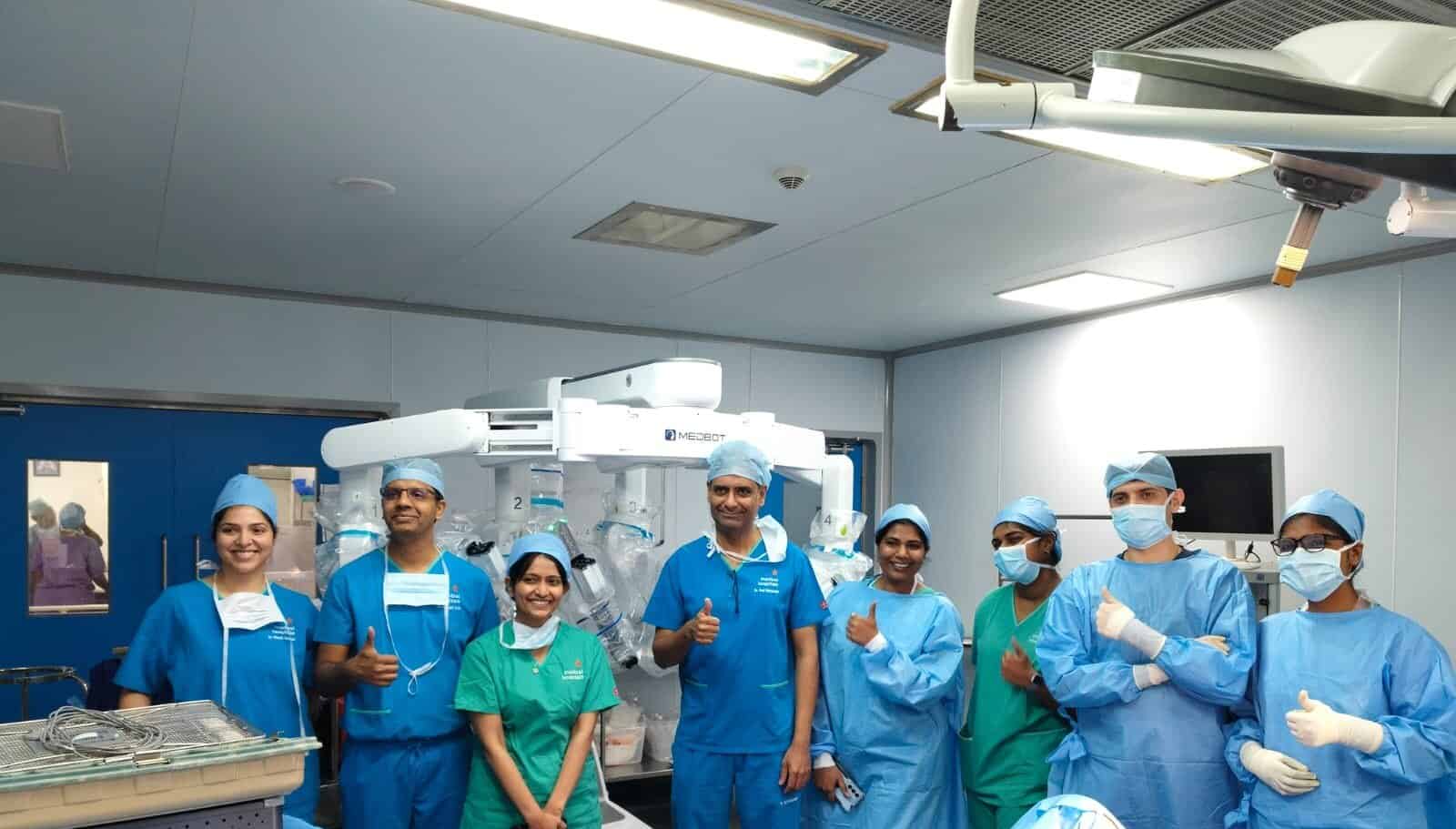
A robotic cancer surgery or laparoscopic cancer surgery approach may be considered only in carefully selected cases—for example, a relatively small, well-defined tumor in a favorable location that can be removed intact without compromising the surgical margin.
Minimally invasive surgery should not be selected merely because the incisions are smaller. If the tumor is large, close to major vessels, or requires removal of multiple organs, attempting keyhole surgery may create unnecessary difficulty or risk.
I am experienced in robotic, laparoscopic, minimally invasive, and open cancer surgery. This allows me to recommend the approach based on the tumor rather than being limited to one technique.
For retroperitoneal sarcoma, cancer clearance and patient safety take priority over the size of the incision.
You may read more about Minimally Invasive Cancer Surgery, Robotic Cancer Surgery, Laparoscopic Cancer Surgery, and Open Cancer Surgery.

Before surgery, I explain what the operation is intended to achieve, which organs may need to be removed, important risks, possible alternatives, and the expected recovery.
Preparation may include:
After surgery, the patient may initially be monitored in a recovery unit or ICU, depending on the complexity of the operation and overall health.
Pain relief, breathing exercises, early movement, nutrition, blood-clot prevention, and careful monitoring of bowel and kidney function are important parts of recovery.
The hospital stay varies considerably. A smaller operation may require a shorter admission, while multi-organ or blood-vessel surgery may require longer monitoring. Recovery continues after discharge, and strength usually returns gradually over several weeks.
The final surgical specimen is examined by the pathologist. The report tells us the confirmed subtype, grade, tumor size, treatment effect if preoperative therapy was given, and the status of the surgical margins.
I discuss these findings with the patient and family and explain whether any additional treatment is recommended. Long-term follow-up generally includes clinical review and periodic scans because recurrence can occur even after successful surgery.
More guidance is available under Recovery After Cancer Surgery.

There is no single fixed cost for retroperitoneal sarcoma surgery because these operations vary greatly in complexity.
The cost may depend on:
A meaningful estimate can be prepared after the medical records and scans have been reviewed and the likely operation has been outlined. Even then, the final cost may change if the actual findings during surgery are different from those predicted on imaging.
My team aims to explain the expected treatment components so that the patient and family can plan more confidently.

I currently consult at:
These centers provide access to comprehensive cancer care within a well-equipped hospital environment, supporting multidisciplinary treatment, investigations, and post-operative care when required.
Consultation timings and appointment details can be arranged through phone or WhatsApp for convenience.

This page has been written and medically reviewed under the guidance of Dr. Suraj Manjunath, Senior Surgical Oncologist at Apollo Hospitals, Bannerghatta Road, Bangalore.
The information has been reviewed for medical accuracy, patient clarity, and relevance to cancer surgery decision-making. Cancer treatment is always personalized and depends on factors such as the type of cancer, stage or spread of disease, overall health, co-morbidities, test results, and treatment goals.
The information is meant for patient education and should not replace an in-person consultation, diagnosis or personalized treatment plan.
Consultation Locations: Apollo Hospitals, Bannerghatta Road & Sarjapur Road, Bangalore.
Last reviewed: September 2026
